| Current Translational Medicine, ISSN 0000-0000 online, Open Access |
| Article copyright, the authors; Journal compilation copyright, Curr Transl Med and Elmer Press Inc |
| Journal website https://ctm.elmerpub.com |
Review
Volume 1, 2026, e8

Artificial Intelligence in Functional Lumen Imaging Probe Panometry: Toward Automated Functional and Biomechanical Endoscopy
Ahmed Abdallah Salman 
Internal Medicine Department, Faculty of Medicine, Cairo University, Cairo 11956, Egypt
Manuscript submitted June 29, 2026, accepted September 23, 2026, published online October 10, 2026
Short title: Artificial Intelligence in FLIP Panometry
doi: https://doi.org/10.14740/ctm8
| Abstract | ▴Top |
Background: The impedance planimetry-based functional lumen imaging probe (FLIP) is used to quantify luminal diameter, distensibility, and distension-induced motor activity during sedated endoscopy. FLIP panometry has become a useful adjunct to high-resolution manometry for evaluating esophageal motility, the esophagogastric junction (EGJ), the pylorus, and the anorectum. However, its interpretation is technically challenging, operator-dependent, and reliant on evolving classification schemes, limiting its dissemination beyond expert centers. Artificial intelligence (AI) may provide a means to automate and standardize this interpretation, although its application to FLIP remains in its infancy and is fragmented.
Methods: We conducted a selective, non-systematic narrative review. PubMed, Scopus, Web of Science, and Embase were searched from inception to June 2026, with the search updated to September 2026 at the time of revision, using combinations of terms related to FLIP, EndoFLIP, impedance planimetry, panometry, and distensibility, with terms related to AI, machine learning, deep learning, and relevant disease entities. The core search strategy, the eligibility criteria, and the distinction drawn between direct FLIP-based evidence and related indirect evidence are reported in the Methods. Reference lists were manually searched, and peer-reviewed articles with retrievable full text were prioritized. Because the evidence was heterogeneous and predominantly derived from small single-center studies, the findings were synthesized qualitatively rather than quantitatively pooled, and no study-selection flow diagram or formal risk-of-bias appraisal is presented.
Results: Supervised and deep-learning models can classify FLIP panometry patterns of EGJ opening and the contractile response with accuracies approaching those of expert interpreters, distinguish achalasia subtypes, and provide probabilistic diagnoses. Unsupervised representation learning and mechanics-informed models applied directly to FLIP output extend the analysis from pattern recognition toward biomechanical phenotyping, and have been used to display disease progression and to follow patients after treatment. Emerging research links intraprocedural distensibility to surgical and endoscopic outcomes, and the same computational methods are being investigated for the pylorus, anorectum, and pediatric practice. Across studies, datasets are small, derived from a single device manufacturer, and rarely externally validated; almost none are benchmarked against clinical outcomes rather than expert labels. Agreement with expert readers is not, however, equivalent to demonstrated patient benefit.
Conclusions: AI is closely aligned with the principal limitations of FLIP panometry (interpretive complexity and limited access to expertise) and could transform a niche test into a standardized, real-time decision-support tool. Realizing this potential will require multicenter datasets that span acquisition protocols, probe configurations, and successive versions of the panometry classification, external validation with explicit assessment of calibration, transparent models, and prospective, outcome-anchored studies.
Keywords: Artificial intelligence; Machine learning; Functional lumen imaging probe; Impedance planimetry; Esophageal motility
| Introduction | ▴Top |
The evaluation of dysphagia, non-cardiac chest pain, and suspected gastrointestinal motor dysfunction has shifted over the past two decades from inspection toward the quantification of physiology. High-resolution manometry (HRM), interpreted using the Chicago Classification, established a structured, metric-based framework for assessing esophageal motility and remains the reference standard for this purpose [1, 2]. However, manometry measures the intraluminal pressure generated during swallowing rather than the mechanical behavior of the lumen itself, and it requires a dedicated, awake transnasal catheter study that many patients tolerate poorly. A clinically meaningful subset of symptomatic patients has borderline or normal manometric findings despite important underlying disease, leaving a diagnostic gap that pressure topography alone cannot close [1].
Esophageal symptoms are common and consequential. Dysphagia, heartburn, regurgitation, and non-cardiac chest pain account for a substantial proportion of gastroenterology referrals, and conditions such as achalasia, esophagogastric junction (EGJ) outflow obstruction, and eosinophilic esophagitis (EoE) are increasingly recognized as diagnostic algorithms improve [1, 2]. Accurate characterization of these disorders changes management (directing patients toward myotomy, dilation, antireflux therapy, or anti-inflammatory treatment), so the quality and accessibility of functional testing have direct clinical consequences. Against this background, any tool that can make physiological assessment faster, more reproducible, and more widely available is attractive.
The functional lumen imaging probe (FLIP) interrogates a different and complementary aspect of physiology. Using impedance planimetry within a fluid-filled balloon, FLIP measures luminal geometry and its response to controlled distension, generating distensibility metrics and, when the data are displayed over space and time, distension-induced contractile patterns [3, 4]. Because the probe is deployed during sedated endoscopy, a single sedated encounter can provide structural inspection together with a functional assessment of EGJ opening and esophageal body motility, without requiring the patient to undergo a separate awake study [4, 5]. Applied in this manner, commonly termed FLIP panometry, the technique has evolved from a research tool into an accepted component of the esophagologist’s diagnostic armamentarium, and its indications now extend across achalasia, EGJ outflow obstruction, EoE, and disorders of the pylorus and anorectum [5, 6].
Interpretation remains the principal barrier to wider application. A FLIP study requires the reader to integrate luminal geometry, intra-balloon pressure, and dynamic contractile behavior according to classification rules that are themselves still evolving. This synthesis is time-consuming and remains concentrated in a small number of expert centers [6, 7]. Artificial intelligence (AI) has already transformed adjacent areas of gastroenterology, most visibly through real-time polyp detection during colonoscopy, but its adoption in gastrointestinal motility has been comparatively slow, despite the image- and signal-based nature of FLIP output being precisely the type of structured data for which modern machine learning is well suited [7]. The mismatch is striking: a test that generates rich, standardized, quantitative data is bottlenecked at the human-interpretation stage, exactly where computational methods may provide the greatest assistance.
This review examines how AI can be, and is currently being, applied to FLIP and impedance planimetry. We outline the technology and classification framework that make the data computationally tractable; the interpretive bottleneck that motivates automation; the specific computational tasks (classification, probabilistic diagnosis, representation learning, biomechanical modeling, and outcome prediction) that map onto clinical needs; the clinical domains in which these tools are emerging, from the esophagus to the pylorus, anorectum, and pediatric practice; and the barriers and priorities that will determine whether this approach enters routine clinical care (Fig. 1). Recent reviews have separately addressed FLIP panometry itself and the use of AI in manometric diagnosis, and AI applied to FLIP is considered only briefly within each [6, 7]; the same is true of the broader reviews of esophageal symptom evaluation and of AI across gastrointestinal motility that are discussed later in this article. The present review is therefore positioned as a complement to that literature rather than as a first account of the field. What it adds is a FLIP-specific synthesis organized by computational task, an explicit separation of direct FLIP-based evidence from related indirect evidence, and a critical appraisal of how far the available studies still fall short of demonstrating clinical benefit.
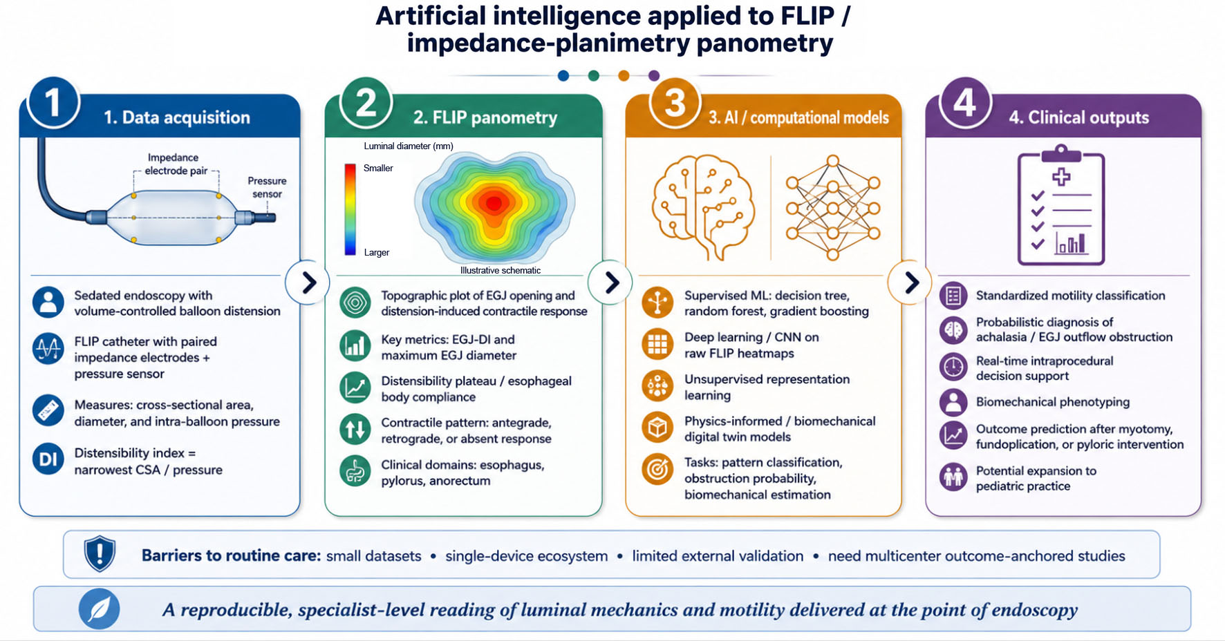 Click for large image | Figure 1. Conceptual pipeline for artificial intelligence applied to functional lumen imaging probe (FLIP) panometry. Volume-controlled balloon distension during sedated endoscopy generates paired cross-sectional-area and intra-balloon-pressure data (impedance planimetry), which are rendered as a FLIP panometry topographic plot of esophagogastric junction opening and the distension-induced contractile response. Computational models (supervised classifiers, deep neural networks, unsupervised representation learning, and physics-informed/biomechanical models) convert these inputs into clinical outputs: standardized motility classification, probabilistic diagnosis, real-time intraprocedural decision support, biomechanical phenotyping, and prediction of treatment outcomes. The heatmap is an illustrative schematic, not patient data. CNN: convolutional neural network; ML: machine learning. |
| Methods | ▴Top |
This is a selective narrative review and is reported as such; it was not conducted as a systematic review, and no study-selection flow diagram, quantitative pooling, or formal risk-of-bias appraisal is presented. PubMed, Scopus, Web of Science, and Embase were searched from inception to June 2026, and the search was re-run and extended to September 2026 at the time of this revision, using the following core strategy: (“functional lumen imaging probe” OR “FLIP” OR “EndoFLIP” OR “impedance planimetry” OR “panometry” OR “distensibility”) AND (“artificial intelligence” OR “machine learning” OR “deep learning” OR “neural network” OR “convolutional” OR “unsupervised” OR “physics-informed” OR “digital twin”). The strategy was then repeated with each of the disease terms achalasia, “esophagogastric junction outflow obstruction,” “eosinophilic esophagitis,” gastroparesis, and “anorectal function” substituted into the second block. Field tags, truncation, and subject headings were adapted to each database, and no language or publication-date restriction was applied. The reference lists of the retrieved primary studies and relevant reviews were manually searched for additional sources.
Consistent with the emphasis on verifiable evidence, peer-reviewed publications with retrievable full text were prioritized. Records were screened by title and abstract, and the remaining reports were assessed in full text against predefined criteria. Eligible reports described the technology or the clinical application of FLIP or impedance planimetry, or the computational analysis of luminal physiology, and provided sufficient methodological detail for the approach to be characterized. Both diagnostic-classification and outcome-prediction applications were eligible, as were studies describing supervised, unsupervised, probabilistic, and physics-informed approaches. Reports were excluded when the full text was not retrievable, when they were conference abstracts or preprints without peer review, when they described neither FLIP nor a computational method transferable to it, or when they duplicated a dataset already represented by a fuller report.
Because the FLIP-specific literature is small, closely related work in HRM and esophageal imaging was retained where the methods are potentially transferable to FLIP. The distinction between the two tiers rests on the data used rather than on the computational method: a report is treated as direct evidence when the model was developed or evaluated on FLIP or impedance-planimetry output, whether the method is supervised, unsupervised, or mechanics-informed, and as indirect evidence when another modality supplied the data. Mechanics-informed modeling of FLIP recordings is therefore direct evidence, whereas the same approach applied to magnetic resonance imaging is not. Indirect reports are identified as such in the text and are tabulated separately from the direct evidence in the study-level evidence table. Statements about what AI can currently achieve with FLIP data rest on the direct evidence alone.
Because the evidence was methodologically heterogeneous, predominantly retrospective, and largely derived from single-center studies, it was synthesized qualitatively. No quantitative pooling, meta-analysis, or formal risk-of-bias assessment was undertaken. Reported performance measures are presented as described by the original authors and are not directly comparable across studies because of differences in reference standards, probe protocols, and patient populations.
| FLIP and the Functional-Biomechanical Paradigm | ▴Top |
FLIP measures luminal dimensions using impedance planimetry. A series of paired electrodes mounted inside a compliant, fluid-filled balloon detects the cross-sectional area at multiple adjacent levels, while a pressure sensor records intra-balloon pressure during stepwise, volume-controlled distension [3, 4]. From these paired measurements, the system derives the technique’s central metric: the distensibility index, calculated as the narrowest cross-sectional area divided by intra-balloon pressure. This metric quantifies how readily a sphincter or luminal segment yields under a given load. It represents a property that neither endoscopic inspection nor pressure topography captures directly and reframes the lumen as a mechanical structure with measurable compliance rather than simply as a conduit [4, 5]. Probes are available in shorter and longer configurations; the 16-cm assembly spans the distal esophagus and EGJ and additionally displays secondary peristalsis as repetitive antegrade or retrograde contractions, allowing the motor response to distension to be evaluated alongside the passive mechanical properties of the wall [5].
In practical terms, a panometry study generates a concise set of metrics. The EGJ distensibility index and maximum EGJ diameter characterize junctional opening; the distensibility plateau and measurements of esophageal body compliance characterize the mechanical behavior of the tubular esophagus; and the pattern of distension-induced contractions characterizes the motor response (Table 1 [4, 5, 8–11]). Normative ranges derived from asymptomatic controls define the boundary between normal and abnormal findings, and successive iterations of the panometry classification have revised category definitions as larger validation cohorts have accumulated. For a machine-learning model, this has an easily overlooked implication: the target labels are not static, and a classifier trained according to one version of the classification scheme must be re-evaluated as the standard evolves.
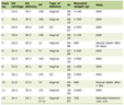 Click to view | Table 1. Common FLIP Panometry Metrics and Their Clinical Interpretation |
Performing the study during endoscopy is the feature that most clearly distinguishes FLIP from manometry operationally, and it has encouraged the concept of “interventional functional diagnostics,” physiological measurement embedded within the therapeutic encounter rather than scheduled as a separate awake catheter study [12]. In practice, the two tests are complementary rather than competing. FLIP characterizes passive tissue properties and distension-induced contractility, whereas HRM characterizes swallow-evoked pressure, and each can detect abnormalities that the other may miss. Their combined use broadens diagnostic yield in esophageal practice, particularly among patients whose symptoms are disproportionate to their manometric findings [13].
The data generated by FLIP panometry are organized into a limited number of clinically meaningful categories, which makes the test tractable for computational analysis. EGJ opening is graded according to the distensibility and maximum diameter achieved during filling, whereas the esophageal body contractile response is classified according to the presence, vigor, and direction of distension-induced contractions. These responses are classified as normal, diminished, disordered, absent, or spastic.
To make these judgments reproducible across operators and centers, expert groups codified acquisition and interpretation in the Dallas Consensus, which specifies how the study should be performed and how EGJ opening and the contractile response should be classified [9, 14]. Iterative validation of the panometry classification in large patient cohorts has refined the category definitions and their thresholds and confirmed their associations with established motility diagnoses [15]. From a machine-learning perspective, this provides an unusually favorable starting point: well-defined inputs (geometry, pressure, and contractile patterns) map onto a constrained set of well-defined output labels, with a published reference standard against which models can be trained and tested. The same standardization that improves human reproducibility also provides the labels, structure, and quality control that automated interpretation depends on [6].
| The Interpretation Bottleneck and the Rationale for AI | ▴Top |
Even with consensus rules, FLIP interpretation is far from trivial. It requires simultaneous reasoning about geometry, pressure, and the timing and direction of contractions, and the resulting judgments are subject to a learning curve and interobserver variability. Agreement between readers is generally good but imperfect, and concordance between FLIP and manometry is incomplete [16]. These disagreements are not merely technical noise. FLIP may reveal abnormal opening or increased wall stiffness when manometric metrics remain within normal limits, and the current Chicago Classification does not fully specify how to reconcile such discordance, leaving clinically relevant phenotypes under-recognized [17]. The practical consequence is a technology whose value is well established at referral centers but is difficult to reproduce in settings with limited panometry expertise.
This is precisely the type of problem for which AI may be suitable, although the analogy with endoscopic image analysis is imperfect. Computer-aided detection during colonoscopy operates on a single, familiar visual stream; FLIP interpretation instead integrates continuous geometric and pressure signals acquired throughout a distension protocol into a categorical physiological diagnosis. The task is therefore closer to time-series and signal classification than to object detection. In gastrointestinal endoscopy more broadly, machine-learning systems can match or exceed human performance on well-circumscribed perceptual tasks. However, translating that success into a tool that generalizes requires representative, multi-institutional data and rigorous external validation, requirements that are easy to state but difficult to meet [18].
Specifically within esophageal physiology, the appeal of AI is fourfold: it can standardize classification across centers, shorten the learning curve, provide rapid results, and, because FLIP is acquired during a procedure, support real-time decision making at the point of care [7, 19]. The goal is not to replace the esophagologist but to make specialist-level interpretation of luminal mechanics available wherever the probe is used [19].
| AI Tasks and Methods Applied to FLIP | ▴Top |
The clinical needs that FLIP must serve map onto a limited set of computational task families, and organizing the literature in this manner clarifies what AI can realistically contribute. Classification addresses the routine diagnostic interpretation; probabilistic modeling addresses uncertainty and triage; representation learning addresses dependence on scarce expert labels and facilitates the discovery of new phenotypes; physics-informed modeling addresses mechanistic interpretation; and outcome prediction addresses the questions that matter most to clinicians and patients. The studies discussed below are grouped accordingly, with the caveat that many systems combine more than one of these functions. Throughout this section a distinction is maintained between studies performed with FLIP or impedance-planimetry data and studies performed with other modalities, principally HRM and magnetic resonance imaging (MRI), which inform method development but do not themselves establish what AI can achieve with FLIP; the direct studies are listed in part A of Table 2 [20–31] and the related studies in other modalities in part B.
Click to view | Table 2. Representative Studies Applying Artificial Intelligence or Computational Modeling to FLIP/Impedance-Planimetry Data (Part A, direct evidence) and to Closely Related Esophageal Physiology Assessed With Other Modalities (Part B, Indirect Evidence) |
Automated classification of panometry patterns
The most developed application of AI to FLIP is the automated classification of panometry studies, and its evolution reflects the broader progression of medical machine learning from engineered features toward learned representations. Early models used supervised decision-tree and random-forest classifiers based on hand-selected descriptors of distension-induced contractility and pressure to identify achalasia subtypes. These models distinguished spastic type III achalasia from non-spastic achalasia with good accuracy, although discrimination among the individual subtypes was more difficult and depended heavily on the quality of the engineered features [20]. That study was retrospective and drawn from a single center, and it was validated internally in a held-out split of the same cohort; the reference standard was the manometric subtype rather than the response to treatment.
Deep learning subsequently shifted the analysis closer to the raw signal. A two-stage convolutional-neural-network pipeline analyzed panometry heatmaps generated from raw FLIP output and achieved 89% accuracy in each binary classification stage against expert labels; it identified 93% of the 28 HRM-confirmed achalasia cases in the test set [21]. This demonstrated that a model could learn clinically relevant structure from minimally processed data rather than relying exclusively on predefined metrics. This model was likewise trained and tested at a single center using a single probe platform, and its reference standard was expert panometry interpretation. These stage-specific accuracies of 89% therefore describe agreement with experienced readers rather than diagnostic correctness measured against an independent clinical endpoint.
The same group has since replaced that convolutional network with a transformer architecture trained to version 2.0 of the panometry classification. In a retrospective single-center cohort of 985 patients divided into training, validation, and test sets without patient overlap, the updated model reached 84% accuracy on the 139-patient test set against 71% for the original, and the reported positive predictive value for normal motility rose from 65% to 91% and that for achalasia from 79% to 84%, although sensitivity for both categories fell slightly and neither model misclassified a normal study as achalasia [22]. Its reference standard was a blinded expert diagnosis derived from HRM using the Chicago Classification version 4.0, supplemented by record review and timed barium esophagram where manometry was inconclusive, which is a more demanding target than expert panometry reading alone. The report is published as a research letter, however, so the methodological detail available for independent appraisal is limited, and the work remains single-center and confined to one probe platform.
The most important recent development has been the move beyond single-center datasets. A multicenter study involving European and North American sites trained several machine-learning classifiers to categorize panometry according to the Dallas Consensus and reported high discrimination for abnormal patterns, EGJ-opening disorders, and contractile-response disorders [23]. Multicenter performance is important because it tests generalizability that single-institution models cannot demonstrate. Alignment with a published consensus standard also ensures that the model outputs remain interpretable to clinicians (Table 2). The advance should not be overstated. The pooled dataset comprised 105 examinations from five centers; the model was trained and tested on a patient-level split within that pooled sample, which is internal validation and not validation in a withheld center; all data were acquired using a single commercial platform; and the target labels were expert Dallas Consensus assignments. Multicenter provenance broadens the case mix but does not by itself demonstrate transportability to a new site.
Probabilistic diagnosis and decision support
Categorical labels, however accurate, discard information about diagnostic confidence. Machine learning can instead provide an estimated probability. A model based on Bayesian additive regression trees estimated the probability of EGJ obstruction from FLIP metrics, achieving an accuracy of 89–90% and an area under the receiver operating characteristic curve of 0.95–0.97, while expressing the result as a probability rather than a binary verdict [24].
The clinical value of this approach is practical. A probability explicitly incorporates uncertainty into decisions made during the procedure, helps triage which patients require confirmatory barium studies or manometry, and allows individual centers to define action thresholds appropriate to their case mix. In a test whose discordance with manometry may itself be clinically informative, communicating uncertainty may be as useful as providing the point estimate. That model was assessed by leave-one-out evaluation together with a holdout test set, both drawn from the same single-center cohort, so its performance has not been examined in an independent center. Two further caveats apply. Discrimination, expressed as the area under the receiver operating characteristic curve, does not establish that the reported probabilities are calibrated, and calibration is the property that matters once a number is used to set an action threshold; calibration was not the focus of the original report and has not been examined in an external cohort. The reference standard also remained an expert-defined diagnosis of obstruction rather than the response to treatment.
Representation learning and unsupervised modeling
Supervised models inherit the limitations and biases of their training labels, and this is a genuine constraint in a field where the reference standard continues to evolve. Unsupervised and self-supervised approaches reduce some of this dependence by learning structure directly from raw physiological data. The most developed example built on FLIP output couples a one-dimensional inverse model, which converts the panometry recording into mechanics-based parameters such as wall stiffness, active relaxation, and contraction pattern, with a variational autoencoder that compresses those parameters into a low-dimensional latent space termed the virtual disease landscape [25]. Patients with motility disorders, EoE, reflux disease, and scleroderma esophagus occupied distinguishable regions of that space, and the same representation was used to display disease progression over time and to estimate the effect of treatment in individual patients.
Because it is derived from FLIP recordings, this work is direct rather than indirect evidence, and it shows that representation learning on panometry can yield quantities that are mechanically interpretable rather than opaque. Its limitations are those of a single-center, retrospective proof-of-concept study: the clusters were compared with existing diagnostic categories rather than with an independent clinical endpoint, and the demonstration of treatment effect was illustrative rather than a prospective test of predictive accuracy. A separate variational autoencoder trained on more than 30,000 raw manometric swallows identified physiologically meaningful contraction patterns without predefined features, with its latent representation encoding contraction strength, propagation, and sphincter relaxation [26]; that model was developed using manometry rather than FLIP and is included as indirect evidence that the same strategy generalizes across modalities. Representation learning offers two principal advantages in either setting: it can reduce dependence on scarce expert labels, and it may identify biomechanical phenotypes overlooked by hand-crafted metrics, thereby refining the classification itself rather than merely automating it.
Physics-informed modeling and the biomechanical digital twin
A distinctive opportunity for FLIP is to combine machine learning with the mechanics of the gastrointestinal wall rather than treating the data as abstract patterns. Mechanics-informed and physics-informed models constrain learning using the governing equations of fluid-structure interaction, ensuring that model outputs remain consistent with the underlying mechanics even when data are sparse. Applied to FLIP itself, this approach has been carried from the virtual disease landscape into a diagnostic framework. In 740 subjects spanning motility disorders and normal physiology, mechanics-based parameters estimated from FLIP were passed through a variational autoencoder and then through linear discriminant analysis to assign each subject a probability of membership in a diagnostic group; the resulting separation of normal EGJ opening from EGJ obstruction aligned closely with both FLIP and manometric diagnoses [27]. An accompanying open software framework has since brought the clinical metrics, the mechanics-based metrics of contraction power and displaced volume, and the virtual-disease-landscape probabilities into a single interface, and reproduced expert categorization of EGJ opening in 121 subjects, with misclassifications mainly falling between adjacent categories and judged acceptable on clinician review [28].
These are the most substantial pieces of direct evidence that FLIP data can support mechanistic rather than purely pattern-based interpretation, and they carry the constraints of the classification literature: both are retrospective and single-center, both take an expert-assigned diagnosis as the reference standard, and neither has been validated in a withheld center or against a prospectively measured treatment outcome. The same family of methods has been applied to esophageal imaging to estimate wall stiffness and active relaxation and to recover information that is not measured directly [29]; that work used MRI rather than FLIP and was a feasibility demonstration in one healthy volunteer, so it indicates what mechanics-constrained learning can recover in principle rather than what it currently delivers for panometry.
FLIP’s paired geometry and pressure data provide a natural substrate for this type of modeling, and the mechanics-based parameter space described above is an early working form of it. The logical endpoint is a patient-specific “biomechanical digital twin” of a sphincter or luminal segment, a mechanistic and interpretable representation of how an individual patient’s tissue behaves under load. Because the outputs are physical quantities with clear clinical meaning rather than opaque scores, this approach may also be easier to validate and trust at the bedside.
Outcome prediction
Ultimately, the endpoint that matters is not agreement with an expert but the clinical outcome, and intraprocedural distensibility is increasingly linked to post-intervention outcomes. Intraoperative measurements obtained during fundoplication suggest that postoperative symptoms are associated with the distensibility achieved across the repair rather than with the specific type of wrap performed, providing a measurable physiological target for surgery [32].
Combining preoperative manometry with intraoperative impedance planimetry may also help identify patients at increased risk of dysphagia after antireflux surgery, allowing counseling and operative technique to be adjusted accordingly [33]. These findings establish the measurable variables on which outcome-focused AI can be developed. Rather than asking only what the current physiological pattern is, models could be trained to predict who will respond to myotomy or dilation, who will have persistent obstruction, and who will develop postoperative dysphagia, questions that are more consequential to patients than the diagnostic label alone. These are observational surgical series rather than studies of AI, and they establish that intraprocedural mechanics carry outcome-relevant information. The mechanics-informed work described above goes a step further, having been used to estimate the effectiveness of treatment and to follow individual patients afterwards within the virtual disease landscape [25]. What is still missing is a model trained and prospectively validated to predict a defined clinical endpoint, such as symptom relief after myotomy or dilation, from FLIP data acquired beforehand.
Cross-modality lessons from manometry
The studies summarized in this section were performed with HRM rather than FLIP and are presented as indirect evidence of methodological feasibility. Because FLIP and HRM share the objective of motility classification, methodological advances in manometric AI offer a readily transferable set of approaches. Deep-learning models for HRM have demonstrated high accuracy in distinguishing normal from disordered motility using architectures designed for spatiotemporal data [30]. A multicenter, multidevice study also showed that machine learning could classify Chicago Classification disorders across different recording systems, demonstrating feasibility in pooled multidevice data, although cross-platform generalizability remains to be established [31].
Forward-looking analyses of esophageal manometry already envisage AI-driven interpretation integrated with FLIP and biomechanical modeling within a single diagnostic pipeline [34]. The substantial overlap in data type, clinical purpose, and even research groups means that techniques validated in manometry (including transfer learning, multimodal fusion, and multidevice training) are natural candidates for adaptation to FLIP panometry.
| Clinical Applications Across the Gut | ▴Top |
Esophagus: achalasia and EGJ outflow obstruction
The esophagus is the region in which both FLIP and its computational analysis are most mature, and the clearest near-term role for AI is to automate tasks that experts already perform well but slowly. FLIP panometry can identify clinically relevant EGJ outflow obstruction consistent with the Chicago Classification and provides validated thresholds for abnormal EGJ opening. This is particularly valuable when manometry or barium studies are equivocal or when symptoms persist despite unremarkable conventional testing [8, 35].
Adjunctive tests such as the timed barium esophagram remain important for confirming outflow obstruction and assessing esophageal emptying [36]. However, obtaining FLIP measurements during the index endoscopy may shorten the diagnostic pathway by combining structural inspection and functional assessment in a single sedated session, sometimes establishing a diagnosis before manometry is performed [37]. Automated panometry classification fits directly into this workflow by converting the expert post-processing currently required by the technique into an immediate, standardized interpretation [20–22]. FLIP can also confirm adequate disruption of the EGJ during and immediately after pneumatic dilation or myotomy, a setting in which real-time AI-based distensibility analysis could provide objective intraprocedural feedback to the operator.
Esophagus: EoE
In EoE, FLIP quantifies the fibrostenotic remodeling that drives dysphagia and food impaction, and that endoscopic appearance and biopsy may underestimate. Reduced esophageal body compliance and distensibility are associated with more severe rings and strictures; reduced distensibility has also been linked to food impaction [10]. Composite physiomechanical scores derived from panometry characterize disease severity and have also been associated with treatment response [11].
These measures are continuous, mechanically grounded variables rather than categorical impressions, making them attractive inputs for machine-learning models. Such models could integrate distensibility, compliance, and the contractile response into reproducible severity indices, track remodeling over time, and potentially predict which patients will respond to dietary, pharmacological, or dilation therapy. As disease-modifying treatments for EoE expand, an objective and automatable assessment of esophageal mechanics could become a useful complement to histology.
Treatment monitoring and surveillance
FLIP’s quantitative outputs are useful not only for diagnosis but also for monitoring disease and treatment over time. In EoE, serial measurements of distensibility may help assess the mechanical response to therapy and complement mucosal eosinophil counts [5]. Following myotomy or dilation for achalasia, intraprocedural distensibility provides an objective measure of the extent of EGJ disruption, information that may be relevant to symptom relief [5].
Longitudinal quantitative monitoring is well suited to machine learning, which can model trajectories rather than isolated time points and identify clinically meaningful changes. This is no longer only a prospect: the mechanics-based virtual disease landscape has already been used to display disease progression across serial studies and to follow patients after treatment [25], although in an illustrative rather than a prospectively validated form. Extending such modeling across serial studies is therefore a natural development of FLIP-based follow-up and parallels the surveillance role that automated tools are beginning to assume elsewhere in endoscopy.
Stomach: the pylorus and gastroparesis
Beyond the esophagus, pyloric EndoFLIP has reshaped the evaluation of gastroparesis by quantifying pyloric distensibility and characterizing the pyloric dysfunction associated with delayed gastric emptying [38]. Distensibility measurements have been used to guide pyloric balloon dilation in patients with refractory symptoms [39] and to confirm adequate disruption of the pyloric sphincter during gastric peroral endoscopic myotomy (G-POEM) and surgical pyloromyotomy [40, 41].
A recurring limitation, however, is that a single preprocedural measurement of pyloric distensibility inconsistently predicts symptomatic response, whereas the change in distensibility achieved during an intervention may be more informative [42, 43]. This is precisely the type of multivariable, weakly signposted prediction problem for which machine learning may be useful. A model integrating pyloric mechanics with gastric-emptying data, symptom scores, and clinical characteristics may identify candidates for pyloric therapy more reliably than any single threshold. The pylorus therefore represents one of the most promising extensions of AI-assisted FLIP beyond the esophagus, although the supporting evidence remains preliminary.
Anorectum and pan-gastrointestinal applications
Impedance planimetry of the anal canal applies the same biomechanical principles to continence and evacuation by measuring the distensibility of the anal sphincter complex. AI applications in anorectal physiology remain nascent but have been identified as a logical extension of work performed in the esophagus [7].
A systematic review of AI in gastrointestinal motility identified applications spanning the esophagus, gastroduodenum, small and large intestine, and anorectum, with FLIP and manometry among the most extensively studied esophageal targets. The review demonstrated both the breadth of the field and the preliminary nature of most efforts, with few externally validated models in any gastrointestinal region [44]. Because these regions share a common analytical substrate, methods developed in the esophagus, where data are currently most abundant, could potentially be adapted to the pylorus and anorectum as suitable datasets become available.
Pediatrics
Pediatric practice is an emerging frontier in which the appeal of a functional assessment performed during sedation is particularly strong, because awake manometry is often poorly tolerated by children. FLIP and AI are among the technologies expected to reshape pediatric gastrointestinal diagnostics [45].
The principal limitation is the availability of data. Pediatric cohorts are smaller, normative distensibility values are not well established, and models trained in adults cannot be assumed to generalize to children. Pediatric FLIP, therefore, represents both a clear opportunity and a domain that requires dedicated, age-specific datasets before any AI-based tool is deployed.
| Integration With the Wider AI Ecosystem | ▴Top |
FLIP-focused AI does not develop in isolation. Contemporary reviews of benign foregut disease and esophageal symptom evaluation increasingly position EndoFLIP alongside AI-assisted imaging and HRM as components of an integrated diagnostic workflow rather than as isolated tests [46, 47].
The development of more mature forms of endoscopic AI is instructive. Experience with computer-aided detection and characterization of Barrett’s neoplasia (including the introduction of regulatory-cleared systems, expansion into depth assessment and procedural guidance, and increasing attention to generalizability and regulation) illustrates both the achievable performance and the challenges that FLIP-based applications are likely to encounter [48]. Similarly, computer-aided assessment of colonoscopy quality demonstrates how automated metrics can be embedded into routine endoscopy and used for feedback and training [49]. Esophageal and anorectal FLIP programs could adopt a similar model as the technology matures.
| Barriers to Clinical Translation | ▴Top |
Several obstacles separate the current proof-of-concept literature from routine clinical use, most of which stem from the small and concentrated nature of the evidence base. First, datasets are limited in size, predominantly derived from single centers, and generated almost exclusively using devices from a single manufacturer. These factors constrain generalizability and increase the risk of overfitting to local acquisition practices. A qualification is warranted here. Because impedance planimetry is currently available for clinical use through a single commercial platform, a genuinely multivendor dataset cannot be assembled at present, and calls for multidevice data should therefore be understood as calls for datasets that span centers, operators, probe configurations, distension protocols, and successive software and classification versions. Vendor independence remains a longer-term aim that depends on the market rather than on investigators. Broad clinical deployment will therefore require multicenter datasets of this kind together with prospective external validation, as limited multi-institutional studies have begun to demonstrate [18, 23, 44].
Second, almost all models are benchmarked against expert interpretation rather than clinical outcomes. Therefore, even excellent concordance with experts does not establish that AI-guided management improves diagnostic accuracy or patient care. The field has largely validated agreement rather than clinical benefit [7]. The distinction is not semantic. Expert labels are themselves imperfect: interobserver agreement for panometry is good but incomplete, and panometry and manometry disagree in a clinically meaningful minority of patients, so a model that faithfully reproduces expert reading also reproduces its errors and its uncertainty [16, 17]. Demonstrating benefit requires prospective designs in which management is guided by the model output and in which the endpoints are diagnostic yield, time to diagnosis, or symptom outcome after treatment.
Third, the opacity of deep-learning models, the established risk of automation bias, and the practical challenges of integrating real-time inference into busy endoscopy workflows must be addressed before clinicians can reasonably rely on these systems. Interpretable outputs and clear presentation of model confidence will be important for clinical adoption [19, 48]. Several practical requirements follow. Model outputs should be calibrated, and calibration should be reported explicitly rather than inferred from discrimination alone, because a probability used at the bedside must mean what it states. The interface should display model confidence and should make the basis of the output inspectable, since automation bias is strongest when a confident label is presented without context. Clinician override must be straightforward, documented, and free of workflow penalty, and recorded overrides should feed back into evaluation as a source of post-market performance data. Integration must also suit the procedural setting, which means inference within the time available during endoscopy, a display that does not compete with the endoscopic image, and an explicit allocation of responsibility for acting on the output.
Fourth, regulatory approval as medical device software, prospective validation, reimbursement, and clearly defined lines of clinical accountability are prerequisites that the FLIP field has only begun to address, although related fields, such as AI-assisted detection of Barrett’s neoplasia, are further advanced [48]. Reimbursement deserves particular attention, because a tool that adds procedural time without an established payment pathway is unlikely to be adopted outside academic centers, and because the principal value of AI-assisted panometry, namely wider access to specialist-level interpretation, accrues largely to the non-expert settings least able to absorb an unfunded cost.
Finally, label noise is an under-recognized problem. Because the reference standard for FLIP panometry continues to evolve and remains imperfectly concordant with manometry, models trained using consensus classifications inevitably inherit the ambiguities of those labels [44]. This problem also has a moving component. The panometry classification has been revised as validation cohorts have grown, and the Dallas Consensus has since standardized acquisition and interpretation, so a model trained to an earlier version of the scheme may misalign with the current one even if its performance appeared satisfactory when it was developed [9, 15]. Version drift of this kind, together with changes in probe hardware, software, and local case mix, makes post-deployment monitoring a requirement rather than an optional refinement. Deployed systems will need periodic revalidation against contemporaneous expert reading, surveillance for performance decay, an auditable record of the model version and of the inputs used for every clinical output, and an agreed procedure for retraining or withdrawing a model whose performance has drifted. None of these barriers is unique to FLIP, but the small and concentrated nature of the current evidence makes each of them particularly consequential.
| Future Directions | ▴Top |
The most promising near-term direction is multimodal integration. Combining FLIP geometry and contractility with HRM, endoscopic imaging, barium studies, and structured clinical data would enable a single model to reason over the same inputs a specialist considers. This could directly address FLIP-manometry discordance, which current single-modality tools cannot fully resolve [19, 44].
Larger pretrained and foundation-model approaches, adapted from their use elsewhere in endoscopy, may improve robustness and reduce the amount of labeled FLIP data that each new center must provide [31]. Physics-informed and digital-twin methods offer a complementary pathway. By generating interpretable, biomechanically grounded outputs, they may earn clinician trust more readily than opaque classifiers and may generalize more effectively because they are constrained by mechanics rather than by the characteristics of a particular dataset [29].
Real-time intraprocedural inference is the application with the greatest immediate clinical potential. Returning a specialist-level interpretation while the probe remains in place could transform panometry from a post hoc analysis into live decision support [34]. Extending validated esophageal methods to the pylorus and anorectum, developing dedicated pediatric normative datasets, and adopting privacy-preserving collaborative strategies such as federated learning (which enables model training across institutions without transferring protected patient records) could collectively broaden the reach and statistical power of these tools [44]. A shared registry of FLIP studies linked to treatment outcomes, together with prospective evaluation designed around those outcomes, would do more to establish clinical value than further incremental gains in agreement with expert labels.
| Limitations of the Study | ▴Top |
This narrative review has several limitations. It was not conducted as a systematic review or meta-analysis, and no formal risk-of-bias appraisal or quantitative synthesis was performed. Although sources were selected using clearly defined search terms and manual reference searching, the selection process remains susceptible to reviewer bias. Priority was given to published, peer-reviewed articles with retrievable full text, which may have underrepresented proprietary or conference-only work. The current AI-FLIP literature is relatively new, rapidly evolving, limited in size, and dominated by a small number of research groups and a single device platform; therefore, this synthesis reflects an early and uneven evidence base. Reported performance metrics were derived from heterogeneous study designs and reference standards and cannot be directly compared across studies. The direct evidence base is itself small: fewer than ten published studies apply AI or mechanics-informed computational modeling to FLIP data, most of them from a small number of groups and several from the same center, and some of the related material discussed here is indirect evidence drawn from manometry and esophageal imaging, which limits how firmly any FLIP-specific conclusion can be drawn.
| Conclusion | ▴Top |
FLIP panometry has emerged as a powerful approach for quantifying the mechanical and motor behavior of the gastrointestinal tract during endoscopy, but its implementation remains constrained by interpretive complexity and limited access to expertise. AI is particularly well-suited to addressing these limitations: it can standardize classification, express diagnostic uncertainty, recover biomechanical properties, and ultimately predict clinical outcomes, while early studies already approach expert-level performance on core tasks.
Nevertheless, the evidence remains preliminary, with studies generally being small, single-center, single-vendor, and benchmarked against expert interpretation rather than patient outcomes; agreement with expert readers should not be read as evidence of patient benefit. If the field can assemble multicenter datasets that span protocols and successive classification versions, validate models externally and prospectively, develop transparent and workflow-ready systems with calibrated outputs and post-deployment monitoring, and anchor evaluation in clinical endpoints, AI-augmented FLIP could transform a specialized measurement into a widely accessible, real-time form of functional and biomechanical endoscopy. Pursued with appropriate rigor, this trajectory could extend the physiological insights provided by FLIP to the many patients currently evaluated outside specialist centers.
| Clinical Significance | ▴Top |
FLIP panometry provides physiological information during endoscopy that complements HRM, but its interpretive complexity has largely limited its use to expert centers. AI-based interpretation could make specialist-level assessment of luminal mechanics and motility available at the point of care, standardize diagnosis across institutions, and support real-time clinical decision-making.
Responsible translation of this potential will require multicenter validation, transparent models, and evaluation against patient outcomes rather than expert agreement alone. For practicing endoscopists, the most immediate benefits are likely to include faster, more consistent panometry interpretations and clearer identification of studies that require specialist input. For healthcare systems, the broader benefit is the potential for wider and more equitable access to functional esophageal assessment.
| Novelty of the Study | ▴Top |
This review is directed specifically at the application of AI to FLIP and impedance-planimetry panometry. Existing reviews address AI in gastrointestinal motility or in high-resolution manometry, where FLIP is generally considered only in passing; the contribution made here is to place FLIP at the center of the analysis and to consolidate a fragmented and rapidly evolving literature into a task-based framework that clarifies what AI can realistically contribute at each stage of analysis.
The review deliberately extends beyond the esophagus to the pylorus, anorectum, and pediatric practice, and it separates direct FLIP-based evidence from the related work in manometry and imaging that is often cited alongside it, so that the small size of the direct evidence base remains visible rather than obscured. It also highlights two directions that distinguish FLIP from image-only endoscopic AI: the biomechanical “digital twin” enabled by paired geometry-pressure data, for which the mechanics-based parameter space derived from FLIP already provides a working prototype, and the transition from models that merely reproduce expert labels to models validated against clinical outcomes. Finally, it outlines a specific research agenda of multicenter datasets spanning protocols and classification versions, external and prospective validation, calibration and interpretability, post-deployment monitoring, and outcome-anchored endpoints, to guide the field from proof of concept toward dependable clinical application.
Acknowledgments
None to declare.
Financial Disclosure
This research received no specific grant from any funding agency in the public, commercial, or not-for-profit sectors.
Conflict of Interest
The author declares no conflict of interest.
Informed Consent
Not applicable; this review did not involve human participants.
Author Contributions
Ahmed Abdallah Salman is the sole author of this manuscript. He conceived the review, designed and conducted the literature search, screened and selected the sources, extracted and interpreted the data, designed the figure and the tables, drafted the manuscript, revised it critically for important intellectual content, and approved the final version submitted for publication. He accepts responsibility for the integrity and the accuracy of the work.
Data Availability
The author declares that the data supporting the findings of this study are available within the article. No new data were created or analyzed, as this review is based on previously published literature.
AI Use Declaration
Artificial intelligence tools were used in a limited and clearly defined capacity during preparation of this manuscript. Claude (Anthropic; accessed June to September 2026) was used to assist with literature searching and with language editing of author-written text, and ChatGPT (OpenAI; accessed June to September 2026) was used to render Figure 1 from a layout and content specification written by the author. No artificial intelligence tool was used to generate scientific content, to design or interpret any analysis, to select or summarize the cited studies, or to formulate any of the conclusions. Every reference was retrieved and verified by the author against PubMed, and all text was reviewed, edited, and approved by the author, who takes full responsibility for the content of the manuscript.
| References | ▴Top |
- Kim JP, Kahrilas PJ. How I approach dysphagia. Curr Gastroenterol Rep. 2019;21(10):49.
doi pubmed - Savarino E, Marabotto E, Bodini G, Furnari M, Della Coletta M, Ghisa M, Barberio B, et al. Advancements in the use of manometry and impedance testing for esophageal functional disorders. Expert Rev Gastroenterol Hepatol. 2019;13(5):425-435.
doi pubmed - Clarke JO, Ahuja NK, Fernandez-Becker NQ, Gregersen H, Kamal AN, Khan A, Lynch KL, et al. The functional lumen imaging probe in gastrointestinal disorders: the past, present, and future. Ann N Y Acad Sci. 2020;1482(1):16-25.
doi pubmed - Dorsey YC, Posner S, Patel A. Esophageal functional lumen imaging probe (FLIP): How can FLIP enhance your clinical practice? Dig Dis Sci. 2020;65(9):2473-2482.
doi pubmed - Savarino E, di Pietro M, Bredenoord AJ, Carlson DA, Clarke JO, Khan A, Vela MF, et al. Use of the functional lumen imaging probe in clinical esophagology. Am J Gastroenterol. 2020;115(11):1786-1796.
doi pubmed - Fass OZ, Carlson DA. Functional lumen imaging probe panometry. Gastrointest Endosc Clin N Am. 2025;35(3):571-585.
doi pubmed - Fass O, Rogers BD, Gyawali CP. Artificial Intelligence Tools for Improving Manometric Diagnosis of Esophageal Dysmotility. Curr Gastroenterol Rep. 2024;26(4):115-123.
doi pubmed - Carlson DA, Prescott JE, Baumann AJ, Schauer JM, Krause A, Donnan EN, Kou W, et al. Validation of clinically relevant thresholds of esophagogastric junction obstruction using FLIP panometry. Clin Gastroenterol Hepatol. 2022;20(6):e1250-e1262.
doi pubmed - Carlson DA, Pandolfino JE, Yadlapati R, Vela MF, Spechler SJ, Schnoll-Sussman FH, Lynch K, et al. A standardized approach to performing and interpreting functional lumen imaging Probe Panometry for esophageal motility disorders: the Dallas consensus. Gastroenterology. 2025;168(6):1114-1127.e5.
doi pubmed - Moosavi S, Shehata C, Kou W, Hirano I, Gonsalves N, Peterson S, Pandolfino JE, et al. Measuring esophageal compliance using functional lumen imaging probe to assess remodeling in eosinophilic esophagitis. Neurogastroenterol Motil. 2023;35(4):e14525.
doi pubmed - Carlson DA, Hirano I, Gonsalves N, Kahrilas PJ, Araujo IK, Yang M, Tetreault MP, et al. Composite score of physiomechanical esophageal function using functional lumen imaging probe panometry in eosinophilic esophagitis. Gastrointest Endosc. 2024;99(4):499-510.e1.
doi pubmed - Vackova Z, Levenfus I, Pohl D. Interventional functional diagnostics in gastrointestinal endoscopy: Combining diagnostic and therapeutic tools in the endoscopy suite with the functional lumen imaging probe. Curr Opin Pharmacol. 2023;73:102414.
doi pubmed - Ellison A, Nguyen AD, Zhang J, Mendoza R, Davis D, Podgaetz E, Ward M, et al. The broad impact of functional lumen imaging probe panometry in addition to high-resolution manometry in an esophageal clinical practice. Dis Esophagus. 2023;36(3):doac059.
doi pubmed - Nguyen AD. The Dallas Consensus for performing and interpreting functional lumen imaging probe panometry: what it means for the clinician. Curr Opin Gastroenterol. 2026;42(4):275-279.
doi pubmed - Fass OZ, Becherano G, Pandolfino JE, Kou W, Ellison AC, Reddy CA, Nguyen AD, et al. Validation of functional lumen imaging probe panometry esophageal motility classification version 2.0: a study of 805 patients. Neurogastroenterol Motil. 2025;37(12):e70188.
doi pubmed - Chen JW, Khan A, Chokshi RV, Clarke JO, Fass R, Garza JM, Gupta M, et al. Interrater reliability of functional lumen imaging probe panometry and high-resolution manometry for the assessment of esophageal motility disorders. Am J Gastroenterol. 2023;118(8):1334-1343.
doi pubmed - Jung KW, Pandolfino JE. When Manometry and Functional Lumen Imaging Probe Disagree: the current limitations of the Chicago Classification Version 4.0 and Probable Extended Indications of Functional Lumen Imaging Probe. J Neurogastroenterol Motil. 2025;31(3):304-312.
doi pubmed - Antonelli G, Eelbode T, Elsaman T, Sharma M, Bisschops R, Hassan C. Building machine learning models in gastrointestinal endoscopy. Gastrointest Endosc Clin N Am. 2025;35(2):279-290.
doi pubmed - Farah A, Abboud W, Savarino EV, Mari A. Esophageal Intelligence: Implementing artificial intelligence into the diagnostics of esophageal motility and impedance pH Monitoring. Neurogastroenterol Motil. 2025;37(9):e70038.
doi pubmed - Carlson DA, Kou W, Rooney KP, Baumann AJ, Donnan E, Triggs JR, Teitelbaum EN, et al. Achalasia subtypes can be identified with functional luminal imaging probe (FLIP) panometry using a supervised machine learning process. Neurogastroenterol Motil. 2021;33(3):e13932.
doi pubmed - Kou W, Soni P, Klug MW, Etemadi M, Kahrilas PJ, Pandolfino JE, Carlson DA. An artificial intelligence platform provides an accurate interpretation of esophageal motility from Functional Lumen Imaging Probe Panometry studies. Neurogastroenterol Motil. 2023;35(7):e14549.
doi pubmed - Fass OZ, Kristinsdottir K, Kaklamanos E, Soni P, Hudson K, Etemadi M, Pandolfino JE, et al. Improved performance of an updated artificial intelligence model for interpretation of Functional Lumen Imaging Probe Panometry studies. Neurogastroenterol Motil. 2025;37(11):e70120.
doi pubmed - Mascarenhas M, Mendes F, Rala Cordeiro J, Mota J, Martins M, Joao Almeida M, Araujo C, et al. Artificial intelligence and FLIP panometry-automated classification of esophageal motility patterns. J Clin Med. 2026;15(1):401.
doi pubmed - Schauer JM, Kou W, Prescott JE, Kahrilas PJ, Pandolfino JE, Carlson DA. Estimating probability for esophageal obstruction: a diagnostic decision support tool applying machine learning to functional lumen imaging probe panometry. J Neurogastroenterol Motil. 2022;28(4):572-579.
doi pubmed - Halder S, Yamasaki J, Acharya S, Kou W, Elisha G, Carlson DA, Kahrilas PJ, et al. Virtual disease landscape using mechanics-informed machine learning: Application to esophageal disorders. Artif Intell Med. 2022;134:102435.
doi pubmed - Kou W, Carlson DA, Baumann AJ, Donnan E, Luo Y, Pandolfino JE, Etemadi M. A deep-learning-based unsupervised model on esophageal manometry using variational autoencoder. Artif Intell Med. 2021;112:102006.
doi pubmed - Halder S, Yamasaki J, Liu X, Carlson DA, Kou W, Kahrilas PJ, Pandolfino JE, et al. Enhancing Chicago Classification diagnoses with functional lumen imaging probe-mechanics (FLIP-MECH). Neurogastroenterol Motil. 2024;36(8):e14841.
doi pubmed - Halder S, Kou W, Goudie E, Kahrilas PJ, Patankar NA, Carlson DA, Pandolfino JE. A software framework for the functional lumen imaging probe-mechanics (MechView). Neurogastroenterol Motil. 2025;37(2):e14981.
doi pubmed - Halder S, Johnson EM, Yamasaki J, Kahrilas PJ, Markl M, Pandolfino JE, Patankar NA. MRI-MECH: mechanics-informed MRI to estimate esophageal health. Front Physiol. 2023;14:1195067.
doi pubmed - Wang Z, Hou M, Yan L, Dai Y, Yin Y, Liu X. Deep learning for tracing esophageal motility function over time. Comput Methods Programs Biomed. 2021;207:106212.
doi pubmed - Mascarenhas M, Mota J, Rala Cordeiro J, Mendes F, Martins M, Cardoso P, Almeida MJ, et al. Artificial intelligence driven diagnosis of motility patterns in high-resolution esophageal manometry: a multicentric multidevice study. Clin Transl Gastroenterol. 2025;16(12):e00941.
doi pubmed - VanDruff VN, Wang CY, Amundson JR, Kuchta K, Hedberg HM, Ujiki MB. Nissen versus Toupet fundoplication quality-of-life outcomes compared by intraoperative EndoFLIP distensibility index range. Surg Endosc. 2026;40(8):6966-6971.
doi pubmed - Al Asadi H, Najah H, Li Y, Marshall T, Salehi N, Turaga A, Finnerty BM, et al. Determination of causes of post-operative dysphagia after anti-reflux surgery based on intra-operative planimetry. Surg Endosc. 2024;38(10):5623-5633.
doi pubmed - Popa SL, Brata VD, Orasan OH, Chiarioni G, Ismaiel A, Padureanu AM, Dumitrascu DI, et al. Future perspectives in esophageal manometry. World J Gastrointest Surg. 2025;17(12):113546.
doi pubmed - Carlson DA, Schauer JM, Kou W, Kahrilas PJ, Pandolfino JE. Functional lumen imaging probe panometry helps identify clinically relevant esophagogastric junction outflow obstruction per Chicago Classification v4.0. Am J Gastroenterol. 2023;118(1):77-86.
doi pubmed - Fass OZ, Pandolfino JE, Schauer JM, Ganesh N, Farina DA, Lat A, Goudie E, et al. Diagnostic accuracy of timed barium esophagram for achalasia. Gastroenterology. 2025;169(1):63-72.
doi pubmed - Carlson DA, Li M, Fass O, Pitisuttithum P, Goudie E, Kou W, Keswani RN, et al. A combined endoscopy and functional lumen imaging probe panometry approach can expedite diagnosis of esophageal motility disorders. Gastrointest Endosc. 2025;102(6):811-821.e4.
doi pubmed - Sharma A, Coles M, Parkman HP. Gastroparesis in the 2020s: new treatments, new paradigms. Curr Gastroenterol Rep. 2020;22(5):23.
doi pubmed - Jehangir A, Malik Z, Petrov RV, Parkman HP. EndoFLIP and pyloric dilation for gastroparesis symptoms refractory to pyloromyotomy/pyloroplasty. Dig Dis Sci. 2021;66(8):2682-2690.
doi pubmed - Hedberg HM, Carbray J, Ujiki MB. Initial experience with endoscopic pyloromyotomy, with description and video of technique. J Gastrointest Surg. 2019;23(8):1706-1710.
doi pubmed - Attaar M, Su B, Wong HJ, Kuchta K, Denham W, Haggerty S, Linn J, et al. Significant changes in impedance planimetry (EndoFLIP) measurements after peroral pyloromyotomy for delayed gastric emptying. Surg Endosc. 2022;36(2):1536-1543.
doi pubmed - Williams AE, Bhanat E, Seerapu VN, Salvemini J, Howell G, Evans MM, Moremen JR. Gastric peroral pyloromyotomy effectiveness in management of all types of gastroparesis. J Gastrointest Surg. 2025;29(6):102031.
doi pubmed - Verga MC, Mazza S, Azzolini F, Cereatti F, Conti CB, Drago A, Soro S, et al. Gastric per-oral endoscopic myotomy: Indications, technique, results and comparison with surgical approach. World J Gastrointest Surg. 2022;14(1):12-23.
doi pubmed - Muggleston D, O'Grady G, Varghese C, Andrews C. Artificial intelligence in gastrointestinal motility diagnostics: a systematic review. Neurogastroenterol Motil. 2026;38(4):e70310.
doi pubmed - Bitton S, Ng K, Khan MA. Editorial: Pediatric gastroenterology and visualizing the digestive tract. Front Pediatr. 2026;14:1877072.
doi pubmed - Nakata JA, Shemmeri E. Benign foregut surgery. Thorac Surg Clin. 2025;35(4):469-477.
doi pubmed - Farah A, Savarino EV, Abboud W, Tatakis A, Mari A. The contemporary diagnostic approaches to esophageal symptomatology. Cureus. 2025;17(2):e78804.
doi pubmed - Roser DA, Ebigbo A. Future perspective of artificial intelligence diagnostics for early Barrett's neoplasia. Digestion. 2026;107(1):91-102.
doi pubmed - Wittbrodt M, Klug M, Etemadi M, Yang A, Pandolfino JE, Keswani RN. Assessment of colonoscopy skill using machine learning to measure quality: Proof-of-concept and initial validation. Endosc Int Open. 2024;12(7):E849-E853.
doi pubmed
This article is distributed under the terms of the Creative Commons Attribution 4.0 International License (CC BY 4.0), which permits unrestricted use, distribution, and reproduction in any medium, including commercial use, provided the original work is properly cited.
Current Translational Medicine is published by Elmer Press Inc.